Whether flying through hurricanes, diving in urban ports to study coral spawning, or sampling deep ocean currents on oceanographic cruises, summer at NOAA’s Atlantic Oceanographic and Meteorological Laboratory (AOML) is filled with scientific activity. To highlight the broad and impactful research conducted at the lab and the passionate scientists driving that work, AOML launched its inaugural Take Action Photography Competition. Spanning the summer field season, the competition invited researchers to submit photos in two categories: Scientists Take Action and Nature Takes Action.
With nearly 50 impressive entries, selecting the finalists was a challenging task. However, we are excited to showcase the standout images and the talented individuals who creatively captured the essence of their research experiences throughout the season.
A big congratulations and thank you to our winners, finalists, and entrants!
Scientists Take Action
Nature Takes Action
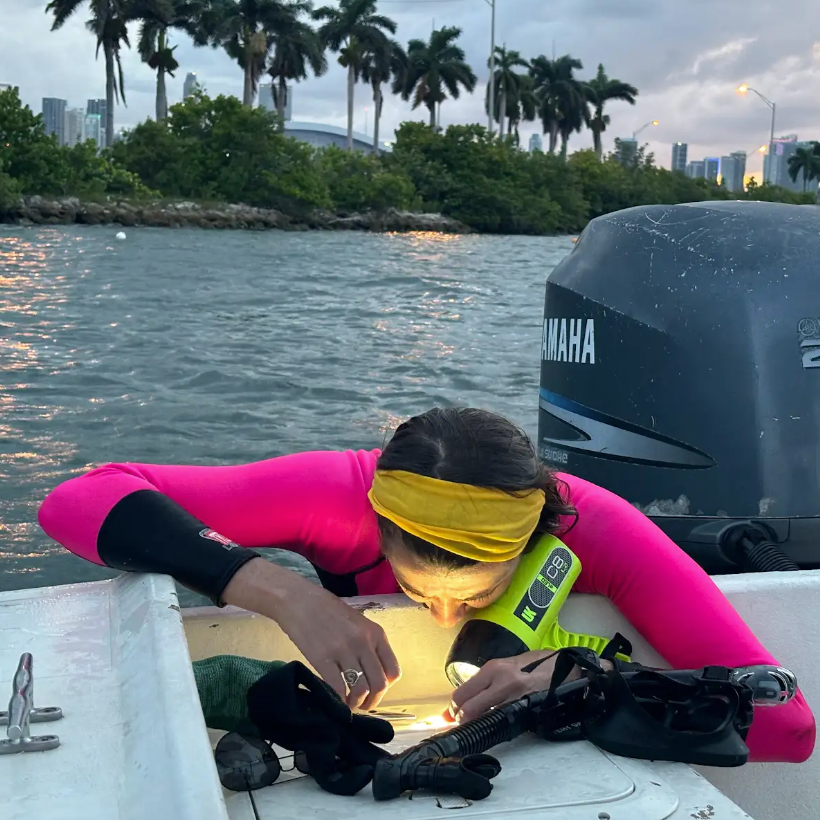
1st place of the Scientists Take Action category and Grand Prize Winner: “Looking for Eggs: by Michael Studivan, Ph.D.
Taking home the grand prize and first place in the Scientists Take Action category is Michael Studivan, Ph.D. with his “Looking for Eggs” photograph. Both Studivan and Rossin are Coral Program researchers who frequently find themselves immersed in field work. In this photograph, Studivan captures Ashley Rossin, Ph.D., checking a coral fragment for eggs at an urban coral site in the Port of Miami. As the grand prize winner, Studivan’s image will be featured as the cover photo on AOML’s FY24 Accomplishment Report.
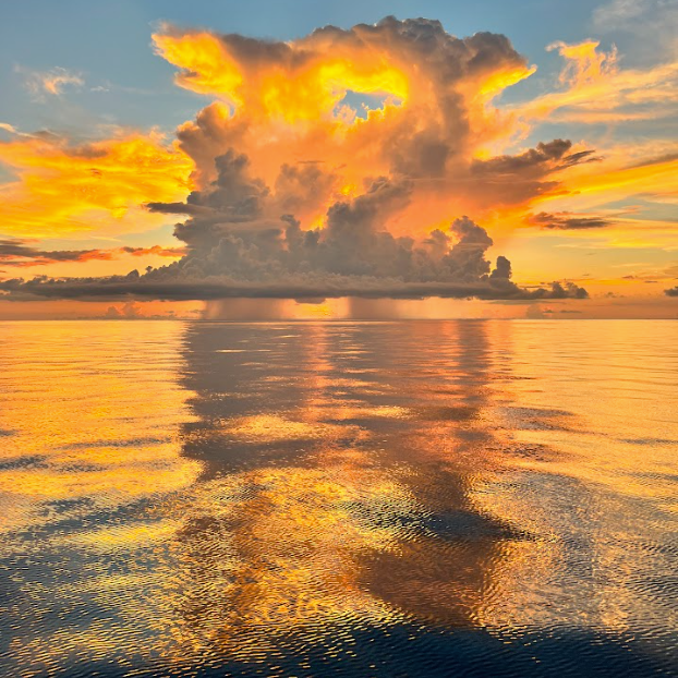
1st place winner of the Nature Takes Action category: “Fire at Sunset” by Tyler Christian
Tyler Christian, a research associate in the Ocean Chemistry and Ecosystem Division, is the first place winner of the Nature Takes Action category. Her photograph, “Fire at Sunset”, was taken on a South Florida Ecosystem Restoration cruise. On these expeditions, researchers collect essential data including hydrographic and biogeochemical variables, carbonate samples, trace metals, eDNA, and plankton tows among other parameters to investigate the status of key marine ecosystems.
Finalists
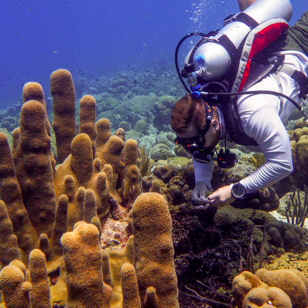
Bradley Weiler, Ph.D., captured images while sampling coral disease in Curacao at a dive site called Double Reef. His photograph entitled, “Diseased Endangered Coral Sampling”, shows the sampling process of the endangered species, Dendrogyra cylindrus, for DNA and RNA sequencing. These coral samples provide critical context and insight needed to compare diseases across species and characterize them from a molecular perspective.
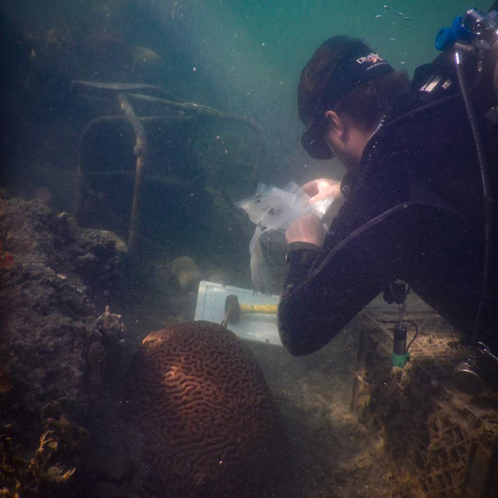
Allyson DeMerlis, Ph.D., submitted photographs from Puerto Rico and the Port of Miami earning her recognition as a finalist. Her photograph called “Urban Coral Sampling” shows Michael Studivan, Ph.D., collecting a coral tissue sample from a Colpophyllia natans coral colony located off of MacArthur Causeway. Note the plethora of marine debris present because of the proximity to the highway.
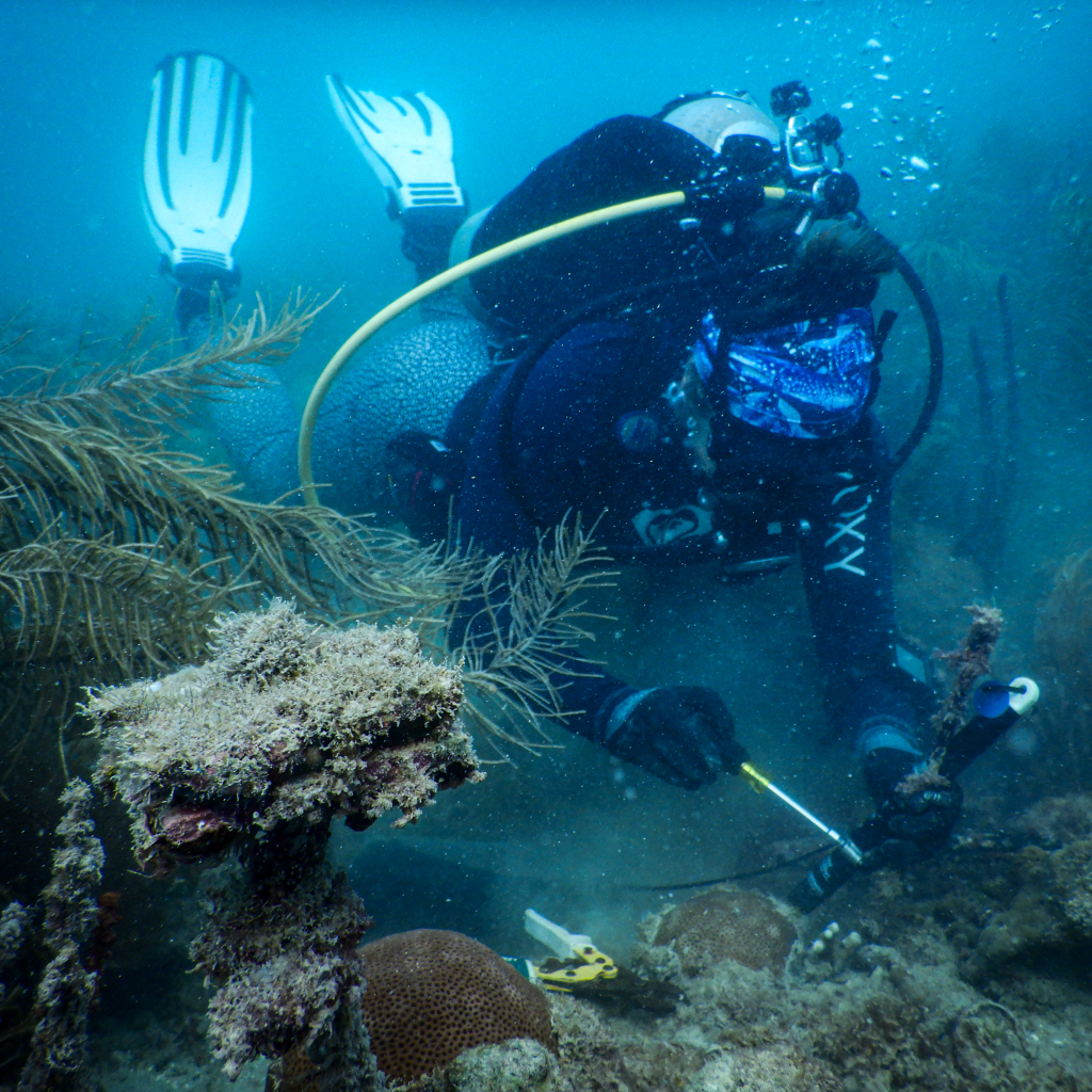
DeMerlis’ photograph titled “Puerto Rico Instruments Deployment” depicts Nikki Besemer collecting a temperature sensor at one of the National Coral Reef Monitoring Program (NCRMP) sites off of La Parguera, Puerto Rico. In the foreground is a tile called a calcification accretion unit (CAU) that measures growth of calcifying organisms.
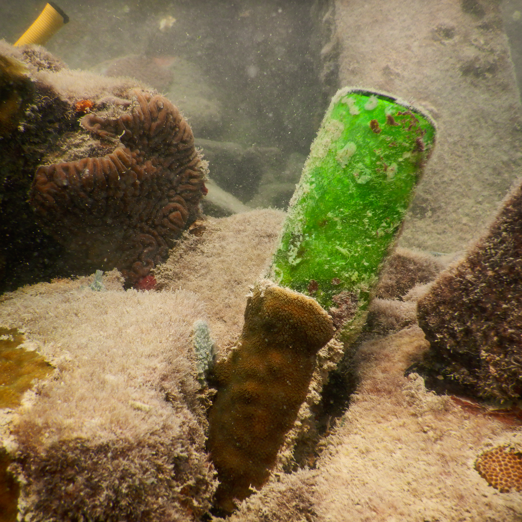
DeMerlis’ “One Coral’s Trash is Another’s Treasure” shows the juxtaposition of coral and urban marine environments. In this unique perspective, corals are growing overtop a discarded glass bottle.
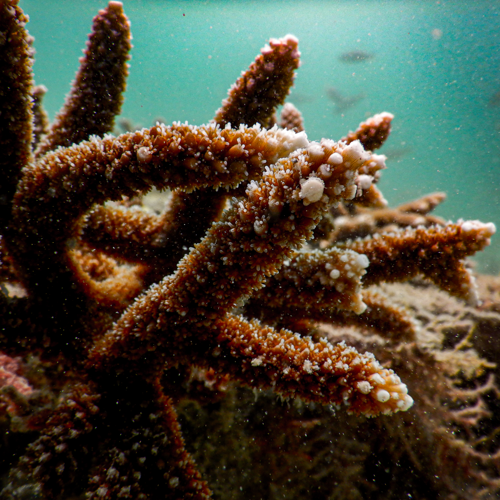
DeMerlis’ “Urban Acropora” photograph depicts staghorn coral with its tentacles extended on the CURES frame at the Coral City Camera site in the Port of Miami.
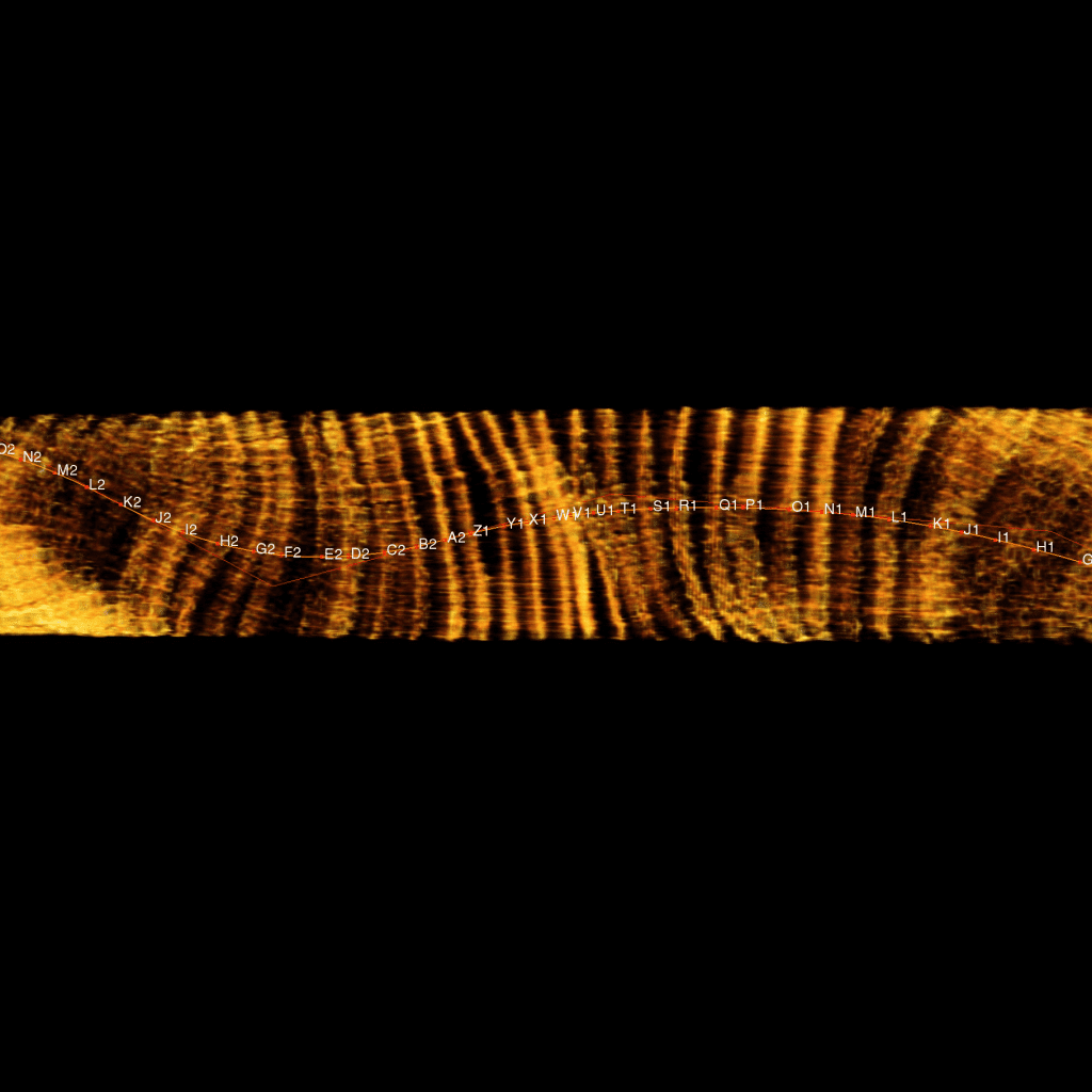
Benjamin Chomitz, Ph.D., is a digital morphology technician with the Ocean Chemistry and Ecosystems Division whose image titled, “Coral Time Machine” shows a CT scan of a coral core from Flower Garden Banks with a line following the growth of the coral and labels for each yearly growth band. Since corals grow by depositing calcified layers, CT scans allow scientists to measure the distance between bands and determine historical growth patterns.
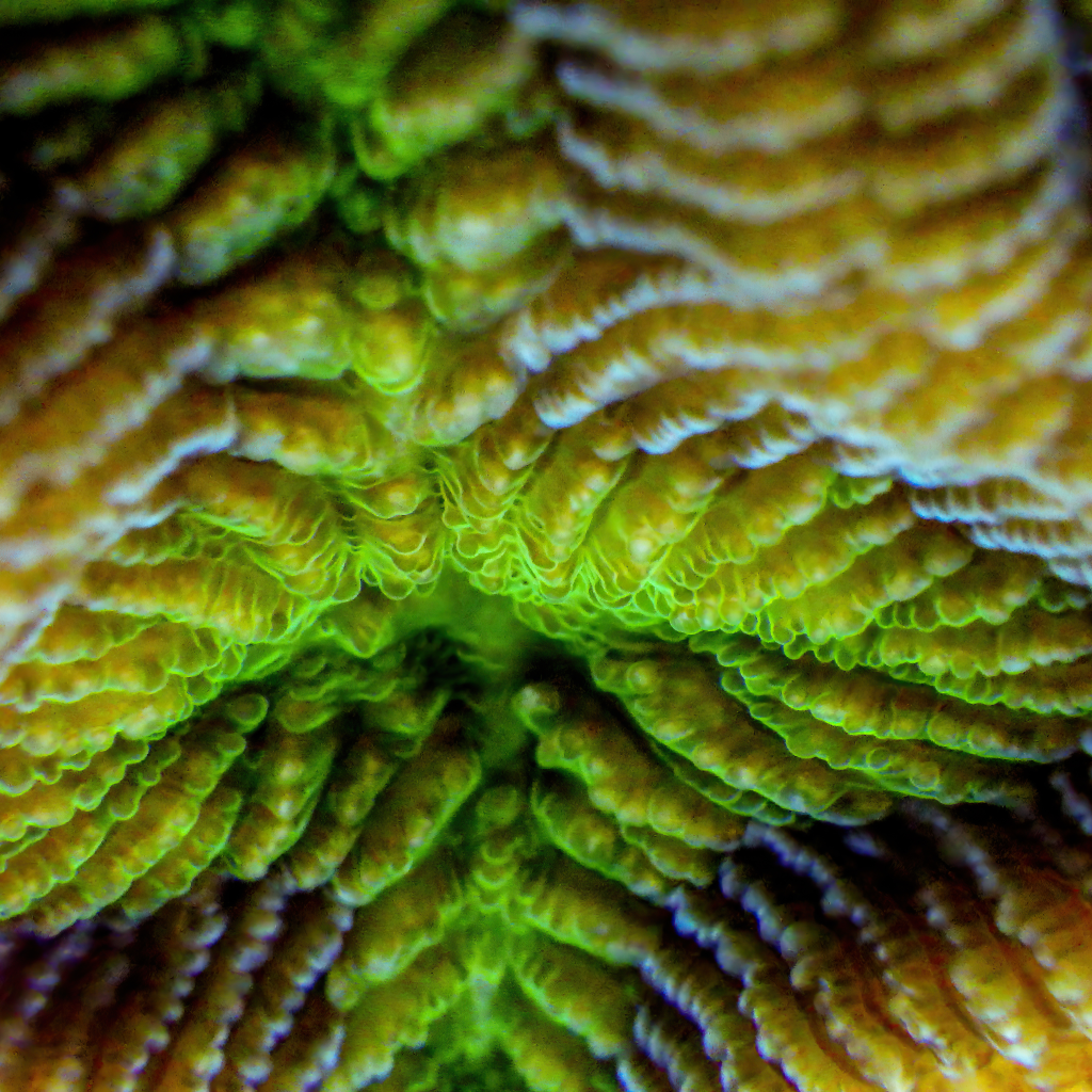
Weiler’s micro-scale photograph of a Colpophyllia natans coral colony has earned him the honor of being a finalist. In this photo, Colpophyllia natans is exhibiting natural fluorescence on the underside of a rocky outcrop near Willemstad, Curaçao.
